Urinary Tract Obstructions: Clinical Case
Urinary Tract Obstruction Case |
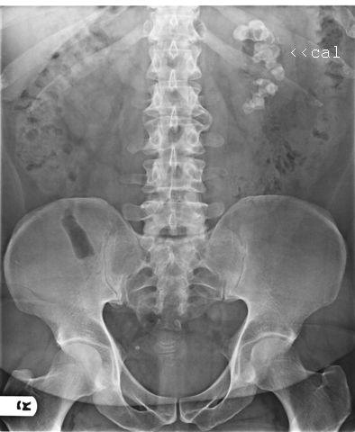
HPI: While in your office, one of your patients, TR, presents to the front desk in severe pain. TR is a 36-year-old Hispanic man who now complains of severe pain in his left flank area that radiates into his left groin. The pain came on suddenly an hour ago and is colicky and sharp/stabbing in nature. He is nauseated and has vomited twice in the last 30 minutes. He denies feeling unwell prior to this episode and denies fever. PMI: TR is an obese man that you recently diagnosed with type II diabetes mellitus that is currently being treated conservatively with diet and exercise. He has been trying to cut-down his coffee intake but continues to drink 6–7 cups per day. He smokes one pack of cigarettes per day and drinks 2–3 beers “after work” daily, more on weekends. He has otherwise been well without previous hospitalizations or surgeries. P/E: TR appears in severe discomfort, is diaphoretic, and is constantly moving, unable to find a comfortable position. Vitals: HR 120; BP 130/87 mm Hg; RR 22; Temp 36.5°C (98°F); SaO2 98% on room air. Head and neck examination is unremarkable. Cardiorespiratory exam reveals tachycardia and tachypnea, but no unusual breath or cardiac sounds. Percussion over his left flank area greatly exacerbates the pain. His abdominal exam is normal. His genital exam is normal without significant testicular discomfort or evidence of hernia. Investigations: TR’s urinalysis reveals frank hematuria. You order an abdominal X-ray series (see Figure 1). |

Figure 1 |
| Question | Your Answer |
What is the likely diagnosis? |
Left-sided nephrolithiasis |
What is the pathophysiology of the pain associated with this condition? |
The kidney stone blocks drainage of the kidney, causing the kidney to expand and stretch the renal capsule. While slow stretching of the capsule may occur with little or no pain (hydronephrosis), acute stretching causes the severe pain associated with nephrolithiasis. |
What are the most common composites of this condition? |
Calcium oxalate makes up 80% of stones. They are 4 times more common in men than women. |
Should you X-ray this patient? |
Since over 90% of stones (calcium, struvite, and cystine) are radiopaque, an abdominal X-ray is a reasonable first radiologic step. Only uric acid stones are radiolucent, so, in a patient with gout or increased cell lysis due to chemotherapy (tumor lysis syndrome), an X-ray is unlikely to be helpful in identifying nephrolithiasis. |
What other investigations can identify the source of this pain? |
Intravenous pyelogram involves injecting a substance that is filtered into the renal tubules and not reabsorbed. It is radiopaque, so subsequent X-rays can chart the path of the substance through the kidneys. Obstruction will lead to either minimal or no uptake into the kidneys and no evidence of flow through the ureter on the affected side. As over 80% of cases are unilateral, the normal side provides a comparison view. |
What investigations help determine the therapeutic course of action? |
The size and location of the stones is important in therapeutic considerations. Small stones usually pass on their own and simply require fluids (oral and/or intravenous, depending on the condition of the patient and level of vomiting) and pain medication. Larger stones that lodge high up in the ureters or calyces are unlikely to pass and require surgical removal or lithotripsy (breaking up of stones, using sound waves). |